Organelle · no membrane
Centrosome
The organiser
In view
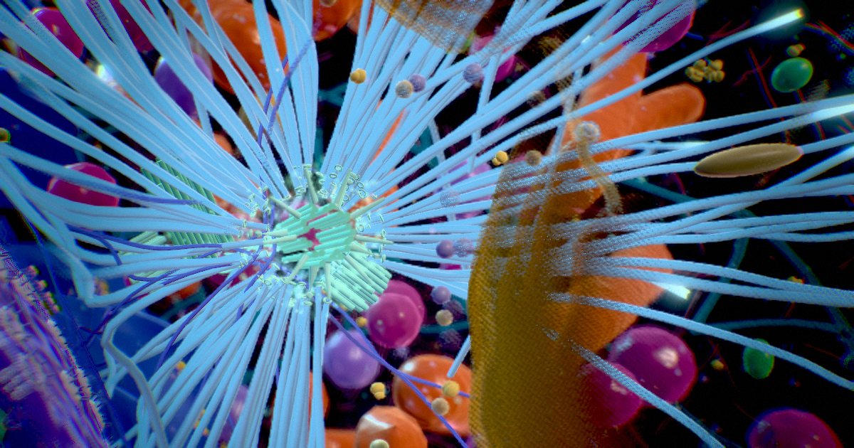
Microtubules sprout from the glowing sleeve around the mother centriole and stream toward the edge of the cell.
The cell’s main microtubule-organising centre. At its heart sit two loosely linked centrioles, each a barrel of nine triplet microtubules arranged like a pinwheel; only the older one carries appendages that hold microtubules. Before division each grows a new centriole at right angles. A layered sleeve of pericentriolar material around the older one launches new microtubules.
It duplicates exactly once per cell cycle, and the two copies become the poles of the mitotic spindle.
- Centriole
- ≈ 250 × 500 nm
- Build
- 9 triplets, pinwheel
- New ones
- Grow at right angles
- Copies
- Once per cell cycle
Did you know?
A centriole can move to the membrane and become a basal body, the base of a cilium that senses the world outside the cell.
Sources
- The Golgi and the centrosome: building a functional partnership · Sütterlin & Colanzi 2010 · J Cell Biol
- Subdiffraction imaging of centrosomes reveals higher-order organizational features of pericentriolar material · Lawo et al. 2012 · Nat Cell Biol
- Centriole structure · Winey & O'Toole 2014 · Philos Trans R Soc Lond B Biol Sci
- Who are you, subdistal appendages of centriole? · Uzbekov & Alieva 2018 · Open Biol
- Once and only once: mechanisms of centriole duplication and their deregulation in disease · Nigg & Holland 2018 · Nat Rev Mol Cell Biol
- Regulating the transition from centriole to basal body · Kobayashi & Dynlacht 2011 · J Cell Biol