Cytoskeleton · 7 nm filaments
Actin filaments
The cell’s muscle
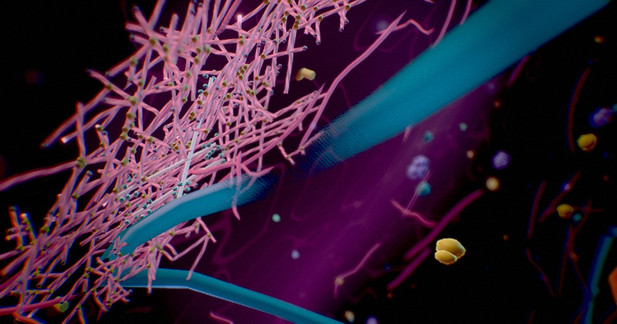
In view
A mesh of short branched filaments and long straight ones lines the inside of the membrane. Up close, new filaments branch off their mothers at about 70° and grow toward the surface, and myosin II tugs on the mesh to keep it taut.
Thin, twisted chains of actin, often the most abundant protein in the cell. Just beneath the membrane they form a dense cortex that gives the surface its shape and strength.
They push out the leading edge when cells crawl, form the cores of microvilli and pinch cells in two at division. Paired with myosin, they power muscle contraction.
- Diameter
- ≈ 7 nm
- Abundance
- ≥ 10 % of protein
- Cortex
- ≈ 0.2 µm thick
- Crawling
- ≈ 0.25 µm/s (fast cells)
Did you know?
When a fish skin cell crawls, every actin filament at its front edge grows by about 100 subunits a second.
Sources
- Monitoring actin cortex thickness in live cells · Clark et al. 2013 · Biophys J
- The interaction of Arp2/3 complex with actin: nucleation, high affinity pointed end capping, and formation of branching networks of filaments · Mullins et al. 1998 · Proc Natl Acad Sci U S A
- Cellular control of cortical actin nucleation · Bovellan et al. 2014 · Curr Biol
- Actin structure and function · Dominguez & Holmes 2011 · Annu Rev Biophys
- How big are the cell’s filaments? · Milo & Phillips · Cell Biology by the Numbers
- Shaping the intestinal brush border · Crawley et al. 2014 · J Cell Biol
- 4.5 The Cytoskeleton · OpenStax · Biology 2e
- Actin and Actin-Binding Proteins · Pollard 2016 · Cold Spring Harb Perspect Biol